Want to learn more? Check out our

MRI findings in IIH

While imaging is performed to assess for secondary causes of intracranial hypertension, there are several imaging findings that can be seen with IIH.

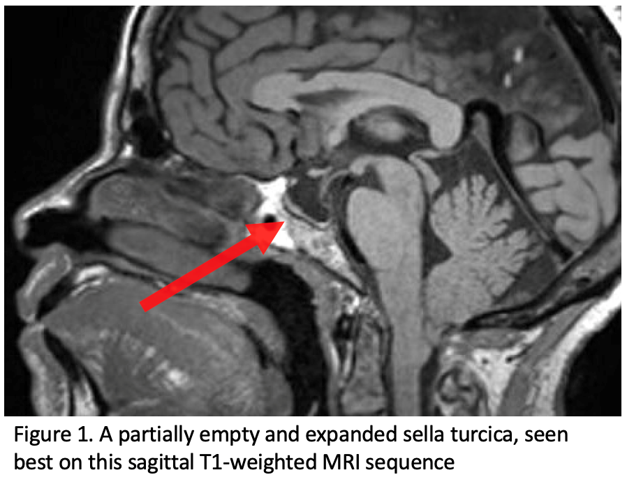
Patients with IIH should get imaging to rule out secondary causes of elevated intracranial pressure, including MR imaging and MR venography of the brain. While imaging is performed to assess for secondary causes of intracranial hypertension, there are several imaging findings that can be seen with IIH. A partially empty sella turcica is a typical feature in the setting of IIH, which appears as a flattened pituitary gland within an expanded sella secondary to chronically increased ICP (Fig 1).

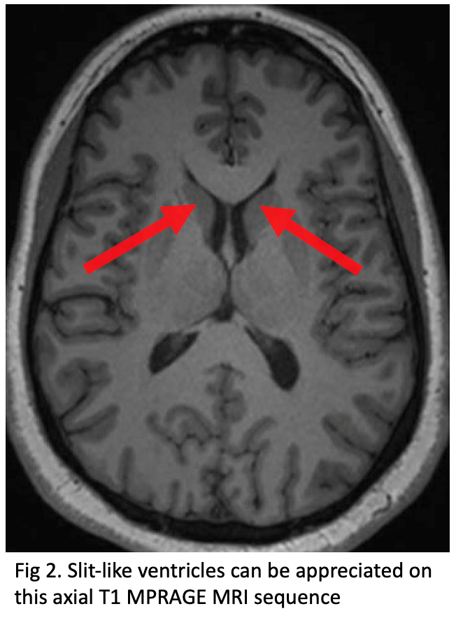
Additionally, slit-like or small compressed ventricles can be seen, typically involving the lateral ventricles (Fig 2).

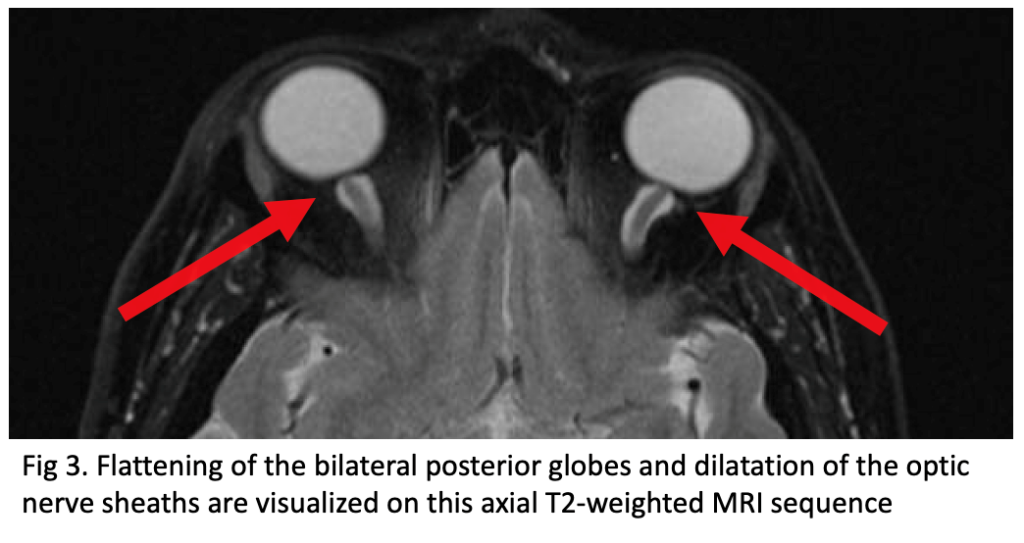
Another set of imaging findings in the setting of IIH is related to the orbits and includes posterior globe flattening, as well as dilatation of the optic nerve sheaths (Fig 3). The optic nerve sheath dilatation appears as increased CSF volume surrounding the intra-orbital optic nerve within the nerve sheath, which can also end up appearing tortuous.


With the use of MR venography, a specialized MR technique that allows non-invasive assessment of the flow within the cranial veins, the major intracranial dural venous sinuses can be well evaluated.

Relatively symmetric dural venous sinus stenosis/narrowing at the transverse and/or sigmoid venous sinuses along the posterior fossa of the intracranial compartment is a classic finding in IIH (Fig 4). An underdeveloped transverse sinus with stenosis of the dominant one can also be found. IIH without any stenosis of the venous sinuses is rare.

In the setting of IIH, the elevated ICP can contribute to prominent arachnoid granulations, also known as Pacchionian granulations, which are projections of the arachnoid membrane into the dural venous sinuses to allow drainage of CSF from the subarachnoid space into the venous system. The prominent arachnoid granulations typically occur along the greater wing of the sphenoid bone and can enlarge to become associated with cephaloceles (Fig 5), which are outward herniations of central nervous system (CNS) contents through a defect in the cranium.

The temporal bone at the skull base is another common location for the development of prominent arachnoid granulations and cephaloceles (Fig 6). Prominent arachnoid granulations can rarely also be associated with CSF leaks through the skull base, for which patients may present with clear rhinorrhea or otorrhea.

Related articles

Omar Choudhri, MD
March 7, 2024
Dr. Omar Choudhri discusses the factors that contribute to the pathophysiology of IIH and advanced imaging to help aid in the diagnosis...
December 14, 2023
Dr. Plenty discusses the importance in understanding how these two conditions are interrelated, as over 95% of people affected with IIH are women ...

Stay Informed

Stay up-to-date on the latest research and resources on this Hub by subscribing to our monthly newsletter

Like what you are seeing?

Join our next webinar.

Thank you! You’re registration is complete for:

IIH Community Chat

Discussion on navigating through the healthcare system

Coming in April